
Radiology
- Home
- Radiology
Medical Ultrasound
Medical Digital X-Ray
Nextcare Diagnostic Radiography produces high quality X-ray for all routine and special X-ray investigations (HSG, IVU, MUG, Barium procedures, etc).
X-Rays make up X-radiation, a form of high-energy electromagnetic radiation.
An X-Ray is a common imaging test that’s been used for decades. It can help your doctor view the inside of your body without having to make an incision. This can help them diagnose, monitor, and treat many medical conditions. Nextcare Diagnostic Centre limited has Digital X-Ray (DR) which provides very high-quality radiography at lowest dose. DR eliminated use of chemicals for film processing. It is fast and produce most accurate result.
In a Digital Radiography, image is produced on a computer monitor immediately.

Doppler/Vascular Ultrasound
Vascular ultrasound uses sound waves (ultrasound machine) to evaluate the body’s circulatory system and help identify blockages in the arteries and veins and detect blood clots.
A Doppler ultrasound study – a technique that evaluates blood flow through a blood vessel – is usually part of this exam.
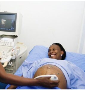
Ultrasound does not use ionizing radiation, has no known harmful effects, and provides images of soft tissues that do not show up on x-ray images.
Our computerised and digitised ultrasound machine is equipped with necessary software and hardware for effective vascular/doppler investigation.
